


Framed or unframed, desk size to sofa size, printed by us in Arizona and Alabama since 2007. Explore now.
Shorpy is funded by you. Patreon contributors get an ad-free experience.
Learn more.


- Baldwin 62303
- Baldwin VO-1000
- Cold
- No expense spared
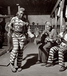
- Tough Guys
- Lost in Toyland
- And without gloves
- If I were a blindfolded time traveler
- Smoke Consumer Also Cooks
- Oh that stove!
- Possibly still there?
- What?!?
- $100 Reward
- Freeze Frame
- Texas Flyer wanted
- Just a Year Too Soon
- WWII -- Replacing men with women at the railroad crossing.
- Yes, Icing
- You kids drive me nuts!
- NOT An Easy Job
- I wonder
- Just add window boxes
- Icing Platform?
- Indiana Harbor Belt abides
- Freezing haze
- Corrections (for those who care)
- C&NW at Nelson
- Fallen Flags
- A dangerous job made worse
- Water Stop
Photos submitted by Shorpy members!
Print Emporium


Radiology: 1920

The halo
of light around the patient's legs looks like he is starting to get that nice radiation glow. Mr. Burns would approve.
Reflection.
The reflection of the doctor on the table near the guy's head is not what it would normally look like.
I can't figure out just how it's looking the way it is.
[It's because the table is concave. Click below to embiggen. - Dave]

Reflection on table
OK you have stumped me. How did the good doctor get a reflection that is not a mirror image, transposed or reversed
Check out the reflection on
Check out the reflection on the table, it's neither of the men pictured.
Memories from A to X
This is very interesting to me. I retired after 35 as a technologist in the field of radiology. When I began my career after two years of training ( 1961-62) I worked with similar equipment. Those old X-ray machines were workhorses for many years. I did not come upon any after 1970 or so.
This picture breaks all the rules -- do not sit out in the room when making an exposure, do not have your assistant stand outside the lead screen. The patient should be propped up on either side a bit. Head turned while in the supine position makes for an unhappy, moving patient.
I have plenty more I could add. However I tend to get a bit long on observation and short of substance.
"How are you feeling?"
"...Like I'm about to get Farked."
Reflection of a reflection
The image of the doctor must be a reflection from the other curved side of the table. It's right side up!
If this contraption didn't
If this contraption didn't kill them, I'm sure something else has by now.
Don't worry sir.
If we can't find the tumor, we'll give you one!
On Shorpy:
Today’s Top 5